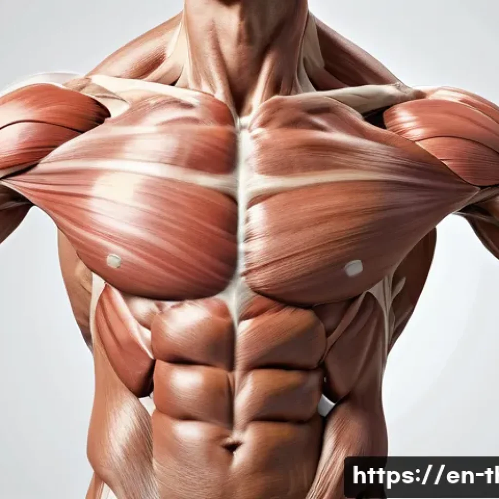
Understanding anatomy is absolutely essential for any physical therapist aiming to provide effective care. It’s the foundation that helps us identify how muscles, bones, and nerves work together to support movement and healing.
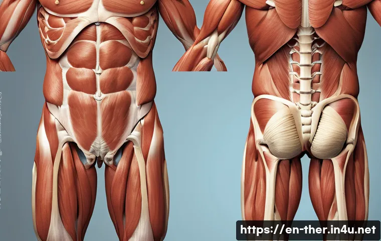
Without a solid grasp of anatomy, designing personalized treatment plans becomes nearly impossible. Over time, I’ve realized that a deep anatomical knowledge not only improves patient outcomes but also boosts confidence in clinical decisions.
Let’s dive into the details and explore why mastering anatomy is a game-changer in physical therapy. Stick with me as we uncover the essentials that every PT should know!
How Musculoskeletal Anatomy Shapes Physical Therapy
Understanding Muscle Function Beyond Names
When I first started out, memorizing muscle names felt like an endless chore, but quickly I realized that knowing what each muscle actually *does* is what really counts.
For example, it’s not just about knowing the biceps brachii flexes the elbow; it’s about understanding how it coordinates with the brachialis and brachioradialis to produce smooth, controlled movements.
This insight changes how you approach strengthening exercises or manual therapy. I’ve found that patients respond better when treatment targets the specific functional role of muscles rather than just the muscle itself.
This functional perspective helps avoid overloading compensatory muscles, which can lead to new pain patterns.
Joint Mechanics and Their Role in Movement
Joints are often underestimated in therapy, but they’re like the hinges on a door—without proper alignment and mobility, everything else struggles. I’ve seen firsthand how subtle restrictions in joint capsules or ligaments can throw off an entire kinetic chain.
Take the shoulder complex, for instance: if the scapulothoracic joint isn’t moving properly, the glenohumeral joint compensates, often leading to impingement or rotator cuff injuries.
That’s why understanding the biomechanics of joints is crucial for crafting interventions that restore natural movement patterns instead of just masking symptoms with pain relief techniques.
The Nervous System’s Influence on Movement Patterns
Muscles and joints don’t work in isolation; they’re constantly guided by the nervous system. Early in my career, I overlooked how nerve injuries or dysfunctions affect muscle activation and coordination.
For example, a patient with a compressed nerve root might present weakness or altered reflexes that mimic muscle pathology, but the root cause lies in nerve conduction.
Recognizing these signs means you can tailor treatments to include neural mobilization or proprioceptive training, which often makes a big difference in recovery speed and quality.
Applying Anatomical Knowledge to Injury Assessment
Pinpointing Pain Origins Through Anatomical Landmarks
One of the trickiest parts of physical therapy is figuring out exactly where pain is coming from, especially when patients describe vague or radiating discomfort.
By using anatomical landmarks, I’ve learned to localize pain sources more accurately. For instance, palpating the greater trochanter helps distinguish between hip joint pathology and referred pain from lumbar spine issues.
This precision guides me in selecting the right diagnostic tests and prevents unnecessary treatments.
Recognizing Patterns in Common Injuries
Anatomy teaches you that certain injuries tend to follow predictable patterns due to biomechanical stresses. For example, the anterior cruciate ligament (ACL) is prone to tears in athletes who perform sudden deceleration or pivoting motions.
Knowing this, I emphasize prevention strategies like neuromuscular training and hip strengthening in at-risk populations. Over time, I’ve seen that clients who understand these patterns take ownership of their rehab and actively engage in exercises that reduce re-injury risk.
Using Palpation and Movement Tests Effectively
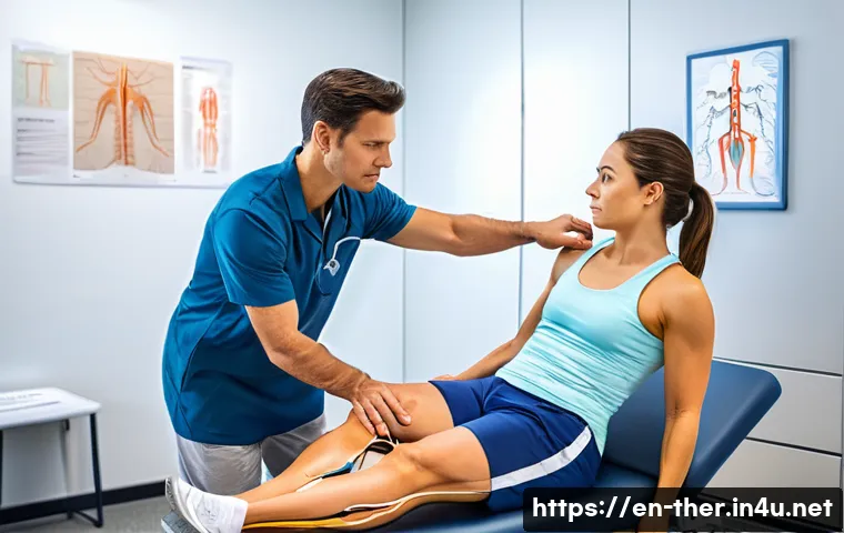
Palpation isn’t just about touching tissues; it’s a diagnostic skill that reveals changes in tissue texture, temperature, and tenderness. When combined with movement tests, it becomes a powerful tool for assessing function.
For example, feeling tightness in the paraspinal muscles while observing limited lumbar flexion can indicate underlying disc issues or muscle guarding.
These findings inform the choice of manual techniques and exercise progressions that I use in practice.
The Critical Role of Neuroanatomy in Treatment Planning
Mapping Dermatomes and Myotomes for Accurate Diagnosis
Neuroanatomy sometimes feels abstract, but mapping dermatomes and myotomes is incredibly practical. I recall a patient who presented with numbness in the thumb and index finger.
By knowing the C6 dermatome distribution, I suspected cervical nerve root involvement rather than peripheral neuropathy. This guided imaging referrals and helped set realistic recovery expectations.
This knowledge also influences the design of sensory re-education and motor control exercises tailored to specific nerve distributions.
Integrating Reflex Testing Into Clinical Assessment
Reflexes provide a window into the integrity of the nervous system. When I first incorporated reflex testing into my routine, I was surprised how much it clarified patient presentations.
For example, an absent Achilles reflex can point to an S1 nerve root lesion, which might not be obvious from strength testing alone. This comprehensive approach reduces misdiagnosis and ensures that therapy addresses the root problem, not just the symptoms.
Neural Tension and Its Impact on Functional Mobility
Neural tension syndromes are often overlooked but can severely limit mobility. I’ve worked with patients whose hamstring tightness wasn’t just muscle stiffness but involved sciatic nerve tension.
Treating neural tension through nerve gliding exercises and positional releases often unlocked improvements that traditional stretching alone couldn’t achieve.
This reinforced for me how essential it is to consider the nervous system’s mechanical properties in treatment.
Understanding the Core’s Anatomy for Stability and Function
Muscles that Make Up the Core and Their Synergy
The core isn’t just the abs—it’s a complex group of muscles including the transverse abdominis, multifidus, diaphragm, and pelvic floor. Early in my career, I treated core weakness by focusing mainly on crunches, but I soon realized that this approach was superficial.
It’s the deep stabilizers that provide true spinal support and coordinate with breathing and pelvic control. I now prioritize exercises that activate these muscles in functional patterns, which leads to better posture and injury prevention.
The Role of Fascia and Connective Tissue
Fascia often flies under the radar, but it’s a crucial component in core stability. I’ve noticed patients with chronic low back pain frequently have fascial restrictions that limit mobility and force compensations elsewhere.
Manual therapy techniques targeting fascial release, combined with active core engagement, have yielded impressive results in my experience. This holistic view of anatomy enhances the effectiveness of rehab programs.
Core Stability’s Influence on Limb Function
Core stability isn’t just about preventing back pain; it’s the foundation for efficient limb movement. For example, during gait or overhead reaching, a strong, well-coordinated core provides a stable base that allows the arms and legs to generate power without undue strain.
I’ve found that when core weakness is addressed, improvements in balance and coordination follow naturally, reducing the risk of falls or overuse injuries.
Integrating Vascular and Lymphatic Anatomy in Rehabilitation
Recognizing Signs of Vascular Compromise
Sometimes symptoms mimic musculoskeletal problems but actually stem from vascular issues. For example, intermittent claudication due to peripheral artery disease can present as leg pain during walking.
By understanding vascular anatomy, I can differentiate these cases through pulse palpation and symptom assessment, ensuring patients get timely referrals to vascular specialists.
Importance of Lymphatic Flow in Post-Injury Recovery
Lymphatic drainage is critical for reducing swelling and promoting tissue healing after injuries. I’ve used manual lymphatic drainage techniques in patients with post-surgical edema or chronic venous insufficiency with great success.
Knowing lymphatic pathways helps tailor these interventions effectively, speeding up recovery and improving comfort.
Enhancing Circulation Through Targeted Exercises
Active movement is one of the best ways to boost circulation and lymph flow. I encourage patients to engage in low-impact aerobic exercises and specific muscle pump activities that stimulate vascular return.
From my experience, this approach not only aids healing but also reduces fatigue and muscle soreness during rehab.
Practical Table: Key Anatomical Structures and Their Clinical Relevance
| Anatomical Structure | Primary Function | Clinical Significance |
|---|---|---|
| Biceps Brachii | Elbow flexion and forearm supination | Common site of tendonitis; important for upper limb strengthening |
| Glenohumeral Joint | Allows wide range of shoulder motion | Prone to instability and impingement; focus of many rehab protocols |
| C6 Dermatome | Skin sensation over thumb and index finger | Used to localize cervical nerve root issues |
| Transverse Abdominis | Core stabilization and spinal support | Key muscle in low back pain rehabilitation |
| Femoral Artery | Supplies blood to lower limb | Assessment site for vascular health; important in peripheral artery disease |
How Anatomical Variations Influence Therapy Outcomes
Recognizing Individual Differences in Anatomy
Not every patient fits textbook descriptions, and I’ve learned that anatomical variations can affect both assessment and treatment. For example, some people have an extra slip of the biceps or variations in nerve branching that can alter symptoms.
Taking time to observe these differences prevents misinterpretation of clinical signs and helps customize therapy.
Adjusting Techniques to Patient-Specific Anatomy
Once you identify an anatomical variation, adapting your techniques is crucial. I recall a patient with a high bifurcation of the sciatic nerve who experienced unusual pain patterns.
Modifying neural mobilization and soft tissue techniques to accommodate this variation made all the difference in her recovery. This experience reinforced the importance of flexibility in clinical reasoning.
Educating Patients About Their Unique Anatomy
Sharing insights about their own anatomy helps patients understand their condition and motivates them to participate actively in rehab. I’ve seen that when patients grasp why certain exercises are prescribed based on their anatomy, compliance improves and outcomes are better.
This also builds trust and reinforces the therapeutic alliance.
Applying Anatomical Insights for Long-Term Injury Prevention
Designing Exercises That Reflect Functional Anatomy
I always aim to prescribe exercises that mirror the way muscles and joints function in real life. For example, instead of isolated quad strengthening, I include multi-joint movements like squats that engage the entire kinetic chain.
This strategy not only builds strength but also improves coordination and reduces the risk of injury during everyday activities.
Incorporating Postural Awareness Based on Structural Anatomy
Posture is deeply connected to anatomy. Understanding spinal curves, pelvic tilt, and scapular positioning helps me guide patients in adopting healthier postures.
Teaching postural awareness has been a game-changer, especially for those with chronic pain or desk jobs, as it addresses root causes rather than just symptoms.
Monitoring and Modifying Activity to Prevent Overuse
Anatomical knowledge helps identify which tissues are vulnerable to overuse. For example, repetitive wrist extension can strain the extensor tendons leading to tennis elbow.
Educating patients about workload balance and early symptom recognition empowers them to adjust activities before minor issues become chronic problems.
This proactive approach saves time and frustration in the long run.
Conclusion
Understanding musculoskeletal anatomy is essential for effective physical therapy. It allows therapists to tailor treatments that address root causes rather than just symptoms. Applying this knowledge improves patient outcomes, speeds recovery, and prevents future injuries. Ultimately, a deep anatomical insight empowers both therapists and patients to work together toward lasting health and function.
Useful Information to Keep in Mind
1. Knowing muscle function beyond names helps target therapy more effectively and prevents compensatory injuries.
2. Joint mechanics play a crucial role in movement patterns, making joint assessment vital in treatment planning.
3. The nervous system influences muscle activation and coordination, so incorporating neuroanatomy enhances diagnostic accuracy.
4. Core stability is foundational for both spinal health and efficient limb movement, emphasizing the importance of deep stabilizer training.
5. Recognizing anatomical variations and educating patients about their unique bodies improves treatment customization and engagement.
Key Takeaways
A thorough grasp of anatomy is not just academic; it directly shapes how therapy is delivered and how patients recover. By integrating knowledge of muscles, joints, nerves, and vascular systems, therapists can create personalized, effective rehabilitation plans. Awareness of individual anatomical differences and functional demands ensures safer, long-term injury prevention. Ultimately, this comprehensive approach fosters better communication, patient trust, and successful outcomes in physical therapy.
Frequently Asked Questions (FAQ) 📖
Q: Why is a strong understanding of anatomy crucial for physical therapists?
A: A solid grasp of anatomy is the backbone of effective physical therapy. It allows therapists to pinpoint exactly how muscles, bones, and nerves interact to produce movement and facilitate healing.
Without this knowledge, it’s like trying to fix a complex machine without understanding how its parts work. From my experience, understanding anatomy deeply leads to more accurate assessments and tailored treatment plans, which ultimately speeds up recovery and improves patient satisfaction.
Q: How does anatomical knowledge impact treatment outcomes in physical therapy?
A: Knowing anatomy inside and out helps physical therapists design interventions that target the root cause of a patient’s problem rather than just alleviating symptoms.
For example, when I encountered a patient with recurring shoulder pain, understanding the intricate muscle attachments and nerve pathways enabled me to create a focused rehab program that addressed underlying imbalances.
This approach not only reduced pain faster but also prevented future injuries, showing how crucial anatomy is for lasting results.
Q: Can mastering anatomy boost a physical therapist’s confidence in clinical decisions?
A: Absolutely. When you fully understand how the body’s structures work together, you feel more confident making clinical decisions because your choices are grounded in solid knowledge.
I’ve noticed that this confidence translates to clearer communication with patients and colleagues, and it helps in adapting treatment plans on the fly when something isn’t working.
Mastering anatomy gives you that professional edge, making you a more effective and trusted therapist.